AI Could Enable Earlier Diagnosis for Brain Tumor Patients
Spanish researchers have developed an image dataset that could revolutionize the diagnosis and treatment of aggressive brain tumors. The breakthrough combines hyperspectral imaging with artificial intelligence.
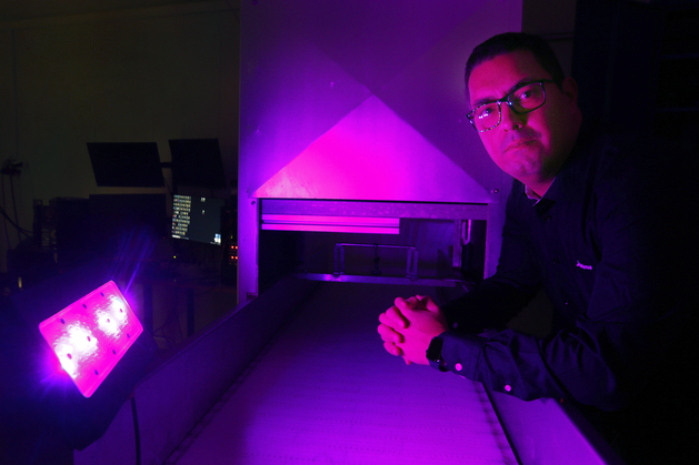
An international research team, led by Samuel Ortega from the Norwegian institute Nofima, has collected 469 hyperspectral images from 13 patients diagnosed with glioblastoma – an aggressive form of brain tumor.
The team has developed a database of advanced brain tumor tissue images that could enable earlier and more precise detection of aggressive cancer cells. Following scientific publication, the database is now available to researchers worldwide.
“By sharing this database openly, researchers can develop better methods for detecting cancer cells in tissue samples. This could lead to faster and more accurate diagnosis,” says Ortega.
The data can be used to train artificial intelligence to identify cancer cells with higher precision than current traditional methods. This could lead to earlier diagnosis, which is crucial for increasing the chances of successful glioblastoma treatment.
International Collaboration
Samuel Ortega is one of several researchers at Nofima’s headquarters in Tromsø working with hyperspectral light and its potential applications. However, the glioblastoma diagnosis work was conducted in Gran Canaria, Ortega’s hometown. The project, funded by Spanish authorities and the EU, is a collaboration between the University of Las Palmas de Gran Canaria, and the Doctor Negrín University Hospital of Gran Canaria.
This is the first hyperspectral imaging dataset published in The Cancer Imaging Archive (TCIA). TCIA is supported by the Cancer Imaging Program (CIP), which is part of the National Cancer Institute (NCI) in the United States. It provides a comprehensive publicly accessible archive of medical images related to cancer.
The images were taken using a hyperspectral microscope that captures both spatial and spectral information, providing unique insight into tumor structure and chemical composition. Patient images were taken at Doctor Negrín University Hospital in Gran Canaria.
Proven Technology
Hyperspectral imaging combines conventional photography with spectroscopy, enabling simultaneous analysis of tissue form and chemical composition. This provides more detailed information than traditional microscope images.
The technology has previously shown promising results in other medical fields, such as early skin cancer detection and blood vessel analysis. It has also been used in food quality control and agriculture. Now it could become an important tool in the fight against brain tumors.
“A major advantage of hyperspectral imaging is its ability to capture details that are difficult to see with conventional microscopy. This can help pathologists make more confident decisions when examining tissue samples,” explains Ortega.
Early Diagnosis Critical
He emphasizes that early diagnosis is critical in fighting this aggressive form of cancer.
“We hope hyperspectral technology this dataset can help save lives by giving doctors a powerful tool for detecting and treating brain tumors at an early stage, and this dataset will contribute to the development of new research in this technology” says Ortega.
HistologyHSI-GB, the first database of its kind for brain tumors, is now freely available to researchers through The Cancer Imaging Archive (TCIA).
Publication
Contact person

Research areas
Quality and measurement methods
Topics
Spectroscopy